Smartgraft is a porcine xenograft supporting a vascularisation of the surgical site through its high porosity and large pores. The bone remodelling and volume stability are supported by the proprietary production process.
Balance high porosity with stable volume remodelling
Cell adhesion
The rough surface of human-like porcine particles facilitates the attachment of new cells.1, 2
Rough surface, large pores, and high porosity enhance bone ingrowth.
Cell migration/infiltration
Facilitates vascularization and bone ingrowth.
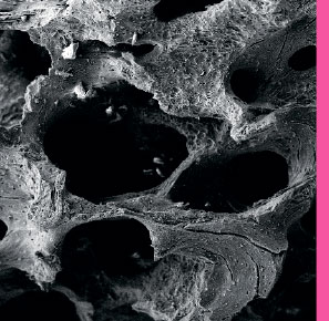
Smartgraft’s high porosity and large pores enhance vascularization, bone ingrowth and osteointegration of the implant after surgery.
Smartgraft’s macropores range from 0.1mm to 1.0mm.
Native porous carbonate apatite possesses the natural pore structure for cell conduction.
Regeneration
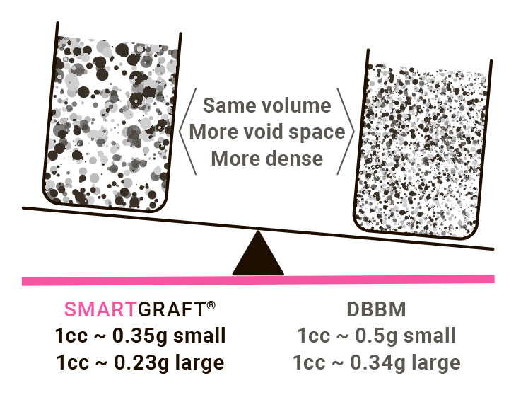
Native porcine graft provides a human-like structure for balanced remodelling.9 The anorganic bone mineral matrix has interconnections that reduce the bulk density of the graft and allows for more void space for the growth of new bones.10
As a porcine-derived bone, Smartgraft accelerates alveolar bone healing as compared to Deproteinized Bovine Bone Mineral (DBBM).11, 12
The proprietary purifi cation process preserves carbonate apatite13, which has been shown to increase the bone forming activities of osteogenic cells and to enhance bioresorption of bone graft by osteoclasts.14-18
The biocompatibility is supported by the proprietary purifi cation process of the graft.19
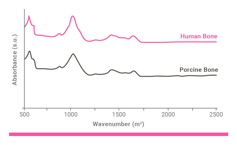
IR Spectra for human and porcine bones
Six reasons to add Hyadent BG to Smartgraft
Sticky bone can be prepared with this out-of-the box gel and Smartgraft in 3 minutes.
As hydrophilic agent, hyaluronic acid (HA) stabilizes blood clot and attracts growth factors to support and accelerate bone formation.20-23
HA supports angiogenesis.24
HA’s high molecular weight reduces swelling and discomfort while supporting scar-less healing.25
HA has natural bacteriostatic properties.26
HA’s special formulation remains present throughout the various phases of the healing process due to its slow degradation pattern (several weeks).22
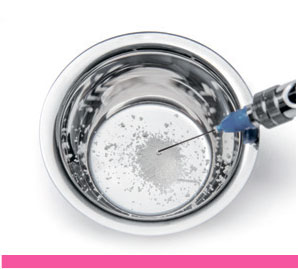
Preparation of stable graft material:
Step 1
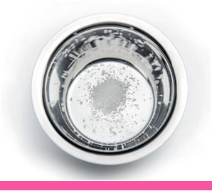
Place bone subtitute granules into a dish.
Hydrate using physiological solution or blood.
Remove any excess fluid.
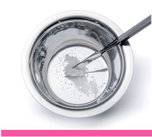
Step 2
Add HYADENT BG to the hydrated bone substitution material.
Step 3
Mix using a spatula.
Repeat steps 2 & 3: Add additional HYADENT BG until the desired consistency is reached (ca. 2/3 Vol% graft material, 1/3 Vol% HYADENT BG).
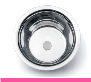
Step 4
Keeping the putty at room temperature for 3-5 minutes may improve the consistency of the putty and make it slightly harder.
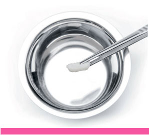
Step 5
Apply putty on to the defect.
Four reasons to use Smartbrane with Smartgraft
Smartbrane ensures adequate tensile strength to safely maintain bone graft stability and structure.27
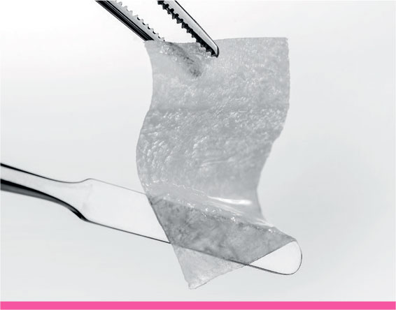
Smartbrane is adaptable to bony surfaces without sticking to the graft or the instrument.31
Smartbrane has a resorption time of 8-12 weeks that can even be extended with Hyadent BG by several weeks.28,29
Smartbrane supports blood clotting and cell attachment.1,3,30
SMARTBRANE rehydrated: excellent adaptation to surfaces without sticking to graft or instrument.
Your regenerative options for your indications
INDICATIONS
SMARTGRAFT
SMARTBRANE
HYADENT BG
Root coverage with CTG
1 x 1.2 ml
Intraosseous defect (1-3 walls) Furcation
0.25 – 1.0 mm granules
15 x 20 mm
1 x 1,2 ml
Fenestration defect
0.5 cc or 1 cc of fine particles
20 x 30 mm
1 x 1.2 ml
Implant dehiscence
0.5 cc or 1 cc of fine particles
15 x 20 mm
1 x 1,2 ml
Extraction socket
1.0 cc of fine particles
10 x 10 mm or 15 x 20 mm
1 x 1.2 ml
Vertical / horizontal augmentation
2.0 cc of large particles
20 x 30 mm or 30 x 40mm
1 x 1,2 ml
Ridge preservation
2.0 cc of large particles
30 x 40mm
1 x 1,2 ml
Sinus floor elevation
2.0 cc of large particles
15 x 20mm / 20 x 30 mm
1 x 1,2 ml
Protection Schneiderian membrane
15 x 20 mm or 20 x 30 mm
1 x 1,2 ml
Видео
Литература
Literature
Deligianni DD, Katsala ND, Koutsoukos PG, Missirlis YF, Effect of Surface Roughness of Hydroxyapatite on Human Bone Marrow Cell Adhesion, Proliferation, Differentiation and Detachment Strength. Elsevier Biomaterials 22 (2001) 87–96
Shu-Thung L et al. (2014) Isolation and Characterization of a Porous Carbonate Apatite From Porcine Cancellous Bone. Science, Technology, Innovation, Aug: 1-13 (data on file)
Frank M. Klenke, Yuelian Liu, Huipin Yuan, Ernst B. Hunziker, Klaus A. Siebenrock, Willy Hofstetter. Impact of Pore Size on the Vascularization and Osseointegration of Ceramic Bone Substitutes in vivo. Journal of Biomedical Materials Research Part A, 2007, 777-786
Hannink G1, Arts JJ. Bioresorbability, porosity and mechanical strength of bone substitutes: what is optimal for bone regeneration? Injury. 2011 Sep;42 Suppl 2:S22-5
Saghiri MA, Asatourian A, Garcia-Godoy F, Sheibani N. The role of angiogenesis in implant dentistry part II: The effect of bone-grafting and barrier membrane materials on angiogenesis. Med Oral Patol Oral Cir Bucal. 2016 Jul 1;21(4):e526-37. doi: 10.4317/medoral.21200. PMID: 27031074; PMCID: PMC4920468.
Data on file
Data on file
Shu-Thung L et al. (2014) Isolation and Characterization of a Porous Carbonate Apatite From Porcine Cancellous Bone. Science, Technology, Innovation, Aug: 1-13 (data on file)
Bracey DN, Seyler TM, Jinnah AH, Lively MO, Willey JS, Smith TL, et al. A decellularized porcine xenograft-derived bone scaffold for clinical use as a bone graft substitute: a critical evaluation of processing and structure. J Funct Biomater. 2018;9(3):45.https://doi.org/10.3390/jfb9030045.
Lai VJ, Michalek JE, Liu Q, Mealey BL. Ridge preservation following tooth extraction using bovine xenograft compared with porcine xenograft: A randomized controlled clinical trial. J Periodontol. 2020 Mar;91(3):361-368. doi: 10.1002/JPER.19-0211. Epub 2019 Aug 23. PMID: 31380563.
Renzo et al.: Tissue Dimensional Changes Following Alveolar Ridge Preservation with Different Xenografts Associated with a Collagen Membrane. Results at the 4-Month Re-Entry Surgery. Int Arch Oral Maxillofac Surg, 2017, 1:003
Guarnieri R, Di Nardo D, Di Giorgio G, Miccoli G, Testarelli L. Effectiveness of Xenograft and Porcine-Derived Resorbable Membrane in Augmentation of Posterior Extraction Sockets with a Severe Wall Defect. A Radiographic/Tomographic Evaluation. J Oral Maxillofac Res. 2019 Mar 31;10(1):e3. doi: 10.5037/jomr.2019.10103. PMID: 31086644; PMCID: PMC6498814.
Method of Preparing Porous Carbonate Apatite from Natural Bone. United States Patent US 8,980,328
F Landi E., Celotti G., Logroscino G., Tampieri A. 2003. Carbonated Hydroxyapatite as Bone Substitute. Journal of the European Ceramic Society 23: 2931–2937.
Spense G., Patel N., Brooks R., Rushton N. 2009. Carbonate Substituted Hydroxyapatite: Resorption by Osteoclasts Modifi es the Osteoblastic Response. Journal of Biomedical Materials Research Part A 217-224.
Doi Y, Shibutani T, Moriwaki Y, Kajimoto T, Iwayama Y. Sintered carbonate apatites as bioresorbable bone substitutes. J Biomed Mater Res 1998;39:603–610
Hasegawa M, Doi Y, Uchida A. Cell-mediated bioresorption of sintered carbonate apatite in rabbits. J Bone Joint Surg [Br] 2003;85:142–147.
Spense G., Patel N., Brooks R., Rushton N. 2009. Carbonate Substituted Hydroxyapatite: Resorption by Osteoclasts Modifi es the Osteoblastic Response. Journal of Biomedical Materials Research Part A 217-224.
Method of Preparing Porous Carbonate Apatite from Natural Bone. United States Patent US 8,980,328.
Muzaffer A et al. ‘The Effect of Hyaluronic Acid-supplemented Bone Graft in Bone Healing: Experimental Study in Rabbits.’J Biomater Appl 2006 20:209
Sasaki T, Watanabe C. ‘Stimulation of osteoinduction in bone wound healing by high-molecular hyaluronic acid.’ Bone. Vol. 16. No.1 January 1995:9-15
Stiller M et al. ‘Performance of β-tricalcium phosphate granules and putty, bone grafting materials after bilateral sinus floor augmentation in humans.’ Biomaterials 2014;35(10):3154-3163.
Mendes RM et al. ‘Sodium hyaluronate accelerates the healing process in tooth sockets of rat.’ Arch Oral Biol 2008; 53:1155–1162
King, S.R., Hickerson, W.L. and Proctor, K.G. (1991) Benefi cial Actions of Exogenous Hyaluronic Acid on Wound Healing. Surgery, 109, 76-86.
Asparuhova M, Kiryak D, Eliezer M, Mihov D, Sculean A. ‘Activity of two hyaluronan preparations on primary human oral fi broblasts’. J Periodontal Res 2018 Sep 27. Epub 2018 Sep 27
Pirnazar P et al. ’Bacteriostatic effects of hyaluronic acid.’ Journal of Periodontology 1999;70:370-374
Internal testing results, data on file.
Internal testing results, data on file.
Eliezer M, Sculean A, Miron RJ, et al. ‘Hyaluronic acid slows down collagen membrane degradation in uncontrolled diabetic rats.’ J Periodontal Res. 2019;00:1–9. https://doi.org/10.1111/jre.12665
Brett D. A Review of Collagen and Collagen-based Wound Dressings. Wounds 2008;20(12).
Data on file
Note: Smartgraft is a registered brand of Regedent AG and manufactured by Collagen Matrix Inc. HYADENT BG is a registered brand and manufactured by BioScience GmbH. Smartbrane is a registered brand and manufactured by Regedent AG
Мы используем файлы cookie для улучшения работы сайта. Продолжая просматривать этот сайт, вы соглашаетесь с условиями использования cookie–файлов. Политика конфиденциальности


 Каталог


 Уведомить
 Купить в 1 клик
 Доставка
 Нет в наличии
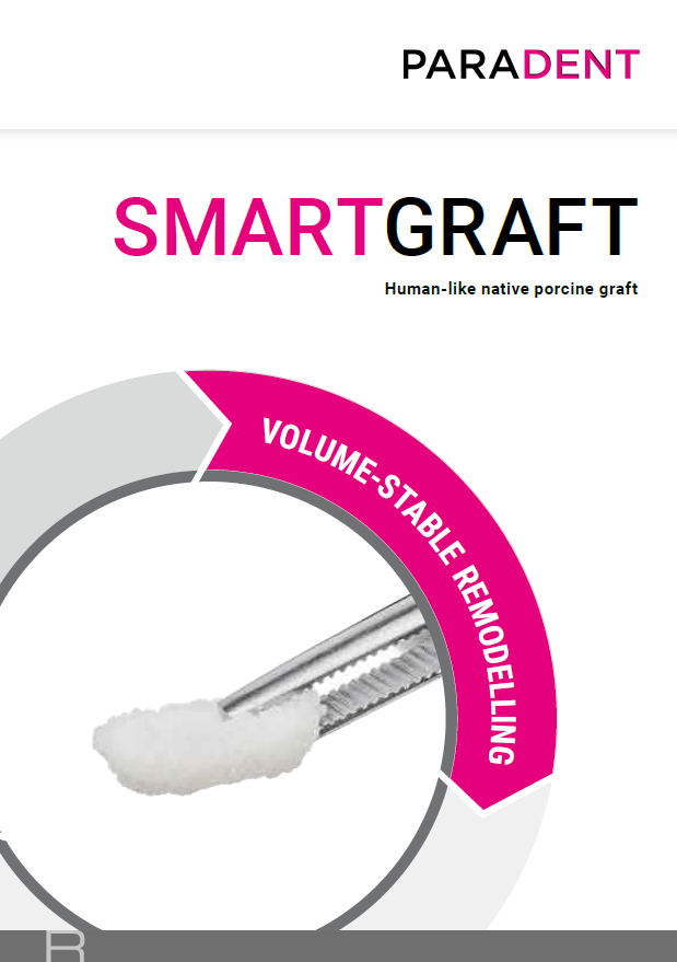

 Подписаться
 Сравнение
 В избранное


