


 Каталог


Case provided By Prof. Dr. Anton Friedman ![]() (Chair and Head Department of Periodontology, School of Dentistry, Faculty of Health, University of Witten, Germany)
(Chair and Head Department of Periodontology, School of Dentistry, Faculty of Health, University of Witten, Germany)
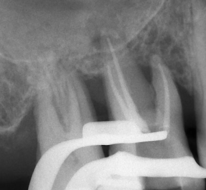
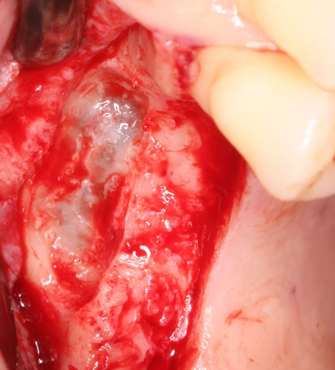
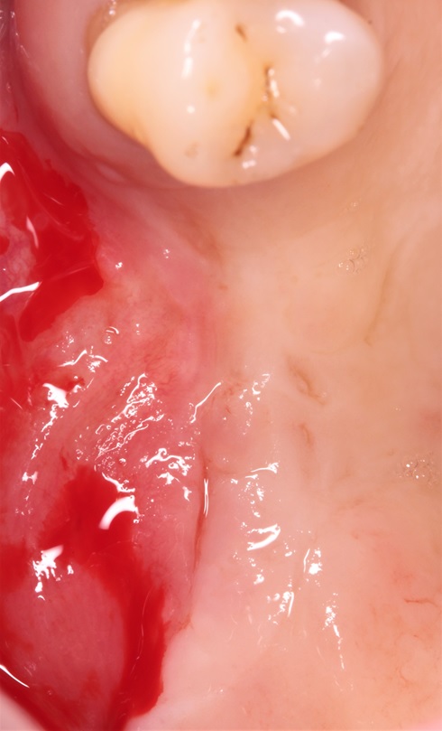
History of hopeless maxillary molars
Residual subantral bone ≈ 2 mm
Lateral window approach ≈ 6 months following tooth loss
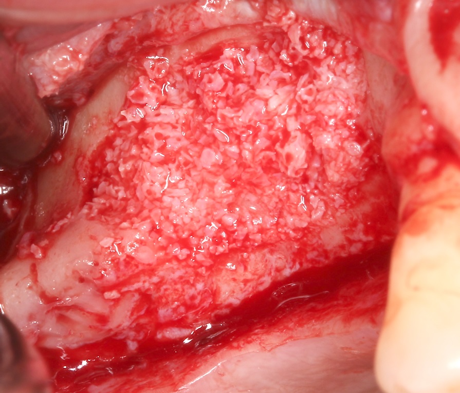
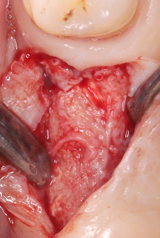
Sinus floor elevation and vertical application of biomaterials
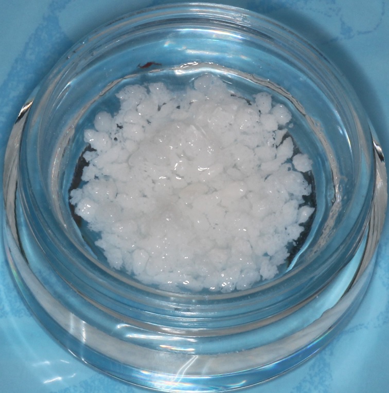
Xenograft (Smartgraft®) hydrated with high weight hyaluronic acid (hyaDENT BG)
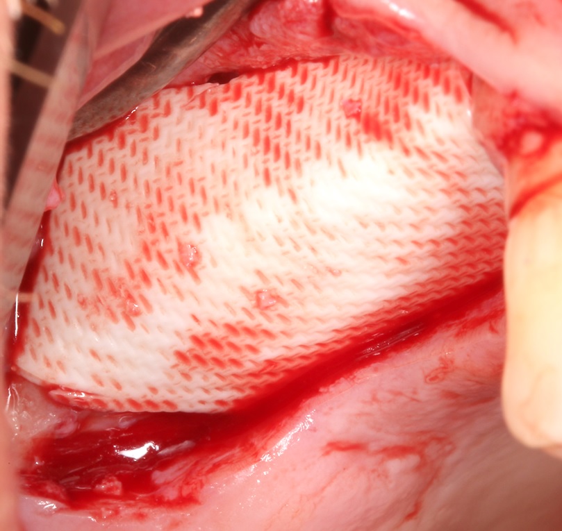
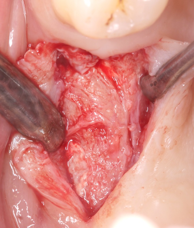
OSSIX®Plus membrane in place covers full extension of the grafted area
Separate membrane fixation unnecessary
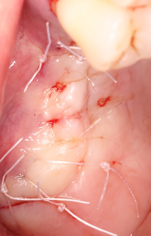
Coronally advanced flap (CAF) and suture
Coronal advancement of the buccal flap (CAF)
Complete tension free closure of the site with PTFE and monocryl sutures
Suture removal ≈ 12 days post-op
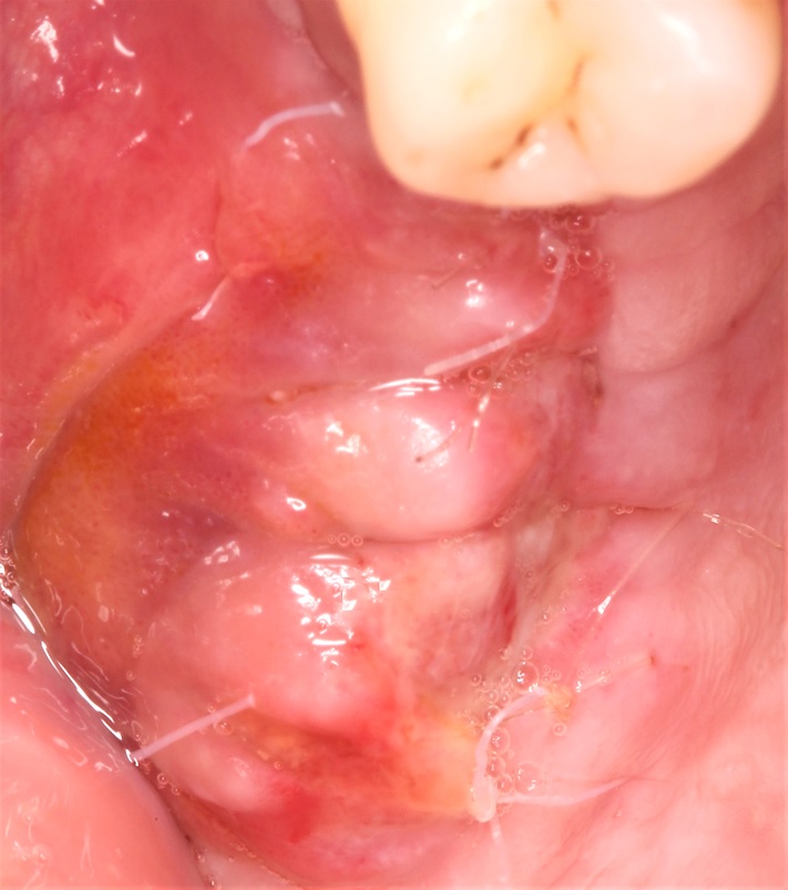
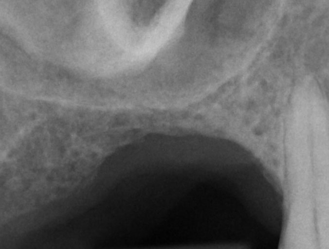
Stage 2 surgery - re-entry and tissue sampling 8 months after SFE
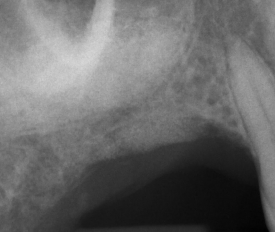
Periapical x-ray displays gain in mineralized tissue at the sinus and supracrestally (arrows)
Soft tissue and ridge dimension at stage 2
Macroscopically mature crestal bone across the ridge
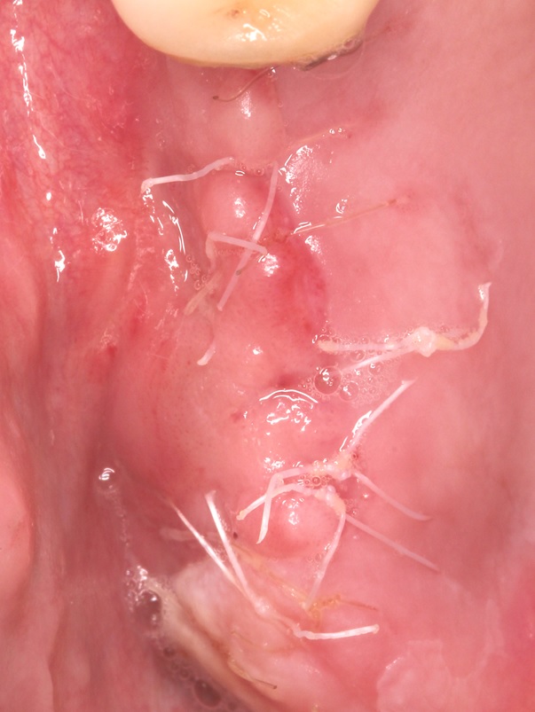
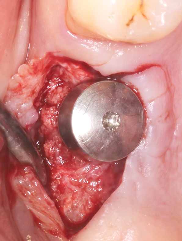
Stage 2 - implant placement 8 months later
Ridge dimension achieved 8 months after SFE and lateral/vertical augmentation
WNI (Straumann SP/WN/10 mm length) in place
Gingiva former in place for transmucosal healing

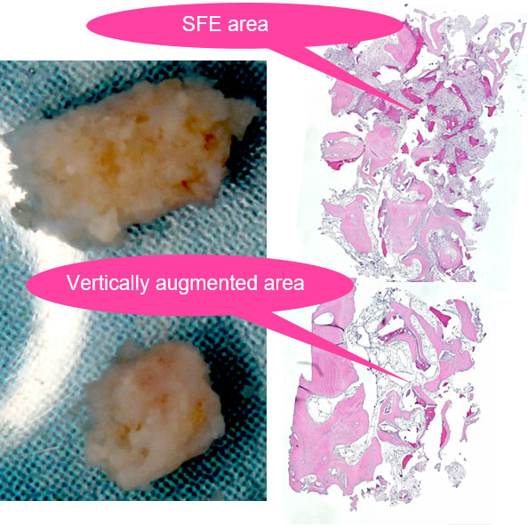
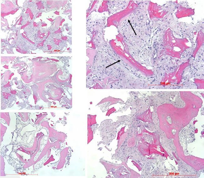
Histological observations in H.E. staining (courtesy Prof. Götz)
Decalcified serial sections reveal various stages of bone formation with appositional bone growth at the residual xenogenic particulate

 Подписаться
 Купить в 1 клик
 Сравнение
 В избранное
 Недоступно

2020-2022 © SIA "Paradent - Baltic". Решения для регенерации костной ткани Латвия, г. Рига, Площадь Республики, 3 – 317, LV-1010 Посмотреть на карте |
+7 925 121-05-02 Email: info@paradent.eu
|